


COMPARISON OF MULTI-STATION WEIGHT BEARING PROPRIOCEPTIVE EXERCISE IN SUBJECTS WITH KNEE OSTEOARTHRITIS
Suraj Kumar*, Ashish Kumar**, V.P. Sharma***
*Associate Professor and HOD, Department of PT at RIMS & R ● E-mail : surajdr2001@yahoo.com
**MPT student, National Institute for Orthopaedically Handicapped, BT Road, Kolkata-90 ● E-mail : ashi_225@yahoo.co.in
*** Professor, Department of Physical Medicine and Rehabilitation, CSM Medical University, Lucknow ● Email : vijaypsharma@yahoo.com
ABSTRACT
Knee osteoarthritis (KOA) is one of the most common diseases which advance with age. It appears no single treatment is sufficient to effectively manage KOA. Diminished strength and proprioceptive acuity are associated risk factors in preventive as well as curative measures. Hence, this investigation was designed to determine the effects of weight bearing proprioceptive exercise in enhancing strength and proprioception in patients with KOA. Randomised trial with concealed allocation and intention-to-treat analysis. A population sample referred in physiotherapy department with diagnosed KOA was included. Variables such as age, sex, height, body weight and body mass index (BMI) was evaluated. Gender wise group distribution was done. Weight bearing proprioceptive training and strengthening exercises was given on alternate day. Treatment session was 6 days a week for 4 week. The data for knee pain, functional disability and proprioceptive acuity were collected and analyzed on the first day and after 4 weeks. There was significant reduction in pain, improved function and proprioceptive acuity in both groups (p< 0.05) but no significant difference between the groups. This study concluded that combined weight bearing Proprioceptive and strengthening exercises are effective in reducing pain and improving function and proprioception in subjects with knee osteoarthritis.
Keywords: Osteoarthritis, proprioception-exercise, strengthening exercise, BMI
INTRODUCTION
Knee osteoarthritis (KOA) is one of the most common disorders, and it is responsible for higher morbidity, particularly in the second half of human life, during which the quality of life is particularly important. Therefore, there is a burden on health from both morbidity and cost (Qing-yu et al., 2006). In contrast to traditional assessments of physical well-being, which have focused largely on the ability of individuals to generate motor output, a greater emphasis is now being placed on sensory feedback acuity. Arguably the most important source of feedback for promoting neural plasticity is our sense of proprioception (Goble, 2010). Sensation is the fundamental ingredient that mediates the proprioceptive mechanism. The articular structures of the body act as sensory chambers which relay proprioceptive information between specific neural pathways within the peripheral nervous system (PNS) and central nervous system (CNS). These neural pathways also transport the necessary sensorimotor information which modulates muscle function (Lephart, 1993). Proprioceptive deficits have been found in KOA (Hewitt, 2002) and it is now considered to play a more significant role than pain in preventing injury in the aetiology of chronic injury and in degenerative joint disease (Lephart, 1995). Clinical aspects of proprioception are measured in tests that measure a subject's ability to detect an externally imposed passive movement, or the ability to reposition a joint to a predetermined position. The initiation of proprioception is the activation of a proprioreceptors in the periphery (Bosco, 2001). The restoration to good proprioception status is widely accepted as a key component in the rehabilitation of other knee, pathologies; improving proprioception in patients with KOA may likely help towards improving knee functions and accelerate the rehabilitation process (Callaghana, 2007).
The primary aim of this study was to evaluate the effect of multi station weight bearing functional pattern exercises and conventional physiotherapy in the management of KOA. The objectives of this study are (i) to find out differences in joint position sense error in male and female before treatment and (ii) to compare the effectiveness of 4 week proprioceptive training in male and female.
METHODOLOGY
A total of 22 patients (11 female and 11 male) who were diagnosed of having KOA by Physicians at OPD, NIOH were recruited for the study. The subjects were screened according to the inclusion criteria and included with intention to treat principle. The inclusion criteria are ages between 40-65 years, radiological/clinical evidence of KOA involving one or both knee joints. Excluded from the study are patients having grade IV KOA on Kellgren-Lawrence Scale, any systemic infection, neurological or; vestibular disorder, deformity of back, hip, knee and ankle, history of either knee trauma during last 3 month or knee surgery, uncontrolled cardiac insufficiency, clinically significant anteroposterior or mediolateral instability of knee, on steroid injection within 6 months in knee joint and uncooperative patient. Informed written consent was taken from all subjects – who participated in the study. The institutional ethical committee, NIOH Kolkata granted approval for the study. The design of the study was pre-post treatment, single blind study randomized control trial. The twenty-two subjects were divided into two groups, Group A (Female) and Group B (male). Both group received conventional physiotherapy in the form of ultrasound therapy and strengthening exercises along with multi station proprioceptive training. The duration of treatment for both groups was 4 weeks. Assessment and treatment was done by one therapist to avoid inter-rater variations. During intervent patients were instructed to avoid either analgesic medication or any other physiotherapy treatment without prior information. The demographic characteristics such as age, weight, height and BMI were taken on the day before receiving treatment. The dependent variables monitored are pain, functional assessment, joint position sense error were measured using NRS, Reduced WOMAC and Electronic Goniometer.
Measurement
Pain was measured using a printed Numerical rating scale (NRS) (AMA, 2010) for every patient on the day before treatment and at the end of four week. Patient were said to mark the number which denotes maximum pain felt in last twenty hour. Functional disability was measured using Reduced Western Ontario and Mcmaster Universities osteoarthritis Index (WOMAC) (Yang, 2007) on the day before treatment and at the end of four week. Patient were said to mark in the column which denotes difficulty in performing the mentioned ADL. Proprioceptive deficit was measured by measuring active joint position sense error (JPS error) (Torres, 2010) on the day before commencement of treatment and at the end of four week. The ability to replicate target knee-joint angles was assessed using an electronic goniometer (Tracker Freedom Wireless Goniometer) (fig.: 1).The test subjects were blindfolded and wore headphones to eliminate visual and auditory cues from the testing apparatus. They wore short nickers to avoid extraneous skin sensation from clothing at the knee area. An Electrogoniometric scale was fastened to the lateral side of the knee with
the fixed arm along the axis of the greater trochanter of femur while the, movable arm was maintained along the axis of lateral malleolus. The goniometer was fastened with 2 Velcro. The Velcro was oriented in a manner so as to minimize skin stretch. A trial was given at each angle before testing. The knee started at full extension. The subject was then asked to flex the knee joint to a pre-determined target angle at 300, 450 and 600. Auditory feedback was provided by therapist during trial at each predetermined angle. Hold up time was 5 seconds at each targeted angle. After returning to the starting position and having remained there for 10 seconds, the subject was asked to flex the knee again to reach the target angle. Three readings were taken at 3 angles. No feedback regarding performance was provided to the subject during the attempt.
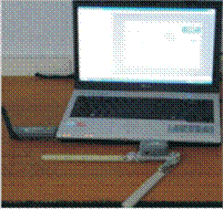
Figure 1, Electronic Goniometer
ngles to be calculated subsequently at base line (before the treatment) marked as JPSO, and at the end of 4th week (after the treatment) marked as JPS .
Intervention
Conventional physiotherapy were in the forms of:
ultrasound therapy (UST):- Frequency 1 MHz, Spatial and Temporal peak intensity of 2.5W/cm2, and pulsed at a duty cycle of 25% for 5 minutes (Huang, 2005) .
Warm up exercises:- (walking, static bicycling and active stretching) of knee, Hip and Ankle each for 2 minutes. Also, included is
Resistive exercise (PRE):- The exercise programs begin with knee flexors and extensors strengthening on Quads table and after that on plinth for hip flexors, hip extensors, hip abductors and hip external rotators. Resistance during knee exercise on quads table was given by appropriate weighted pulley and during hip exercise by appropriate weight cuff. Exercises included 3 sets each of 10 repetitions of open-chain exercises following the Delorme regimen of progressive resistive exercise (PRE) (Bird,2005). Each exercise rhythm of execution was medium slow. The rest period between repetition and sets was 30 seconds and 60 seconds respectively and 5 minute between exercises or legs (Bullock, 2001). Increase in resistance was made gradually every week by 10% increase in weight of previous resistance. Progressions always respect the capacity of adaptation of the subject and exacerbation of discomfort was avoided.
Proprioceptive Training (PrT): PrT programme as formulated by Sekir (2005) was given in the following sequence (Shown in Figure 1):-
- Sit and stand from a standard chair without arm support.
- Drawing two rows of 6 boxes of 50 cm X 50 cm with marker/chalk and was told to walk forward through these 6 boxes “IN-IN-OUT”.
- Walk heel-to-toe along a 3 m line marked on a medium-density polyfoam mat.
- Stair-up and -down a regular 3 steps staircase without hand support.
- Hand side of body. Raise both heels off the ground.
- Foot in same position, Hand clasped behind buttock. And try to hold the position for 10 seconds.
- Hand in same position as in 5, but raise one foot in front and try to hold the position for 10 seconds.
- Foot in same position as in 6, but raise both hand shoulder width apart. Try to maintain the position for
10 seconds. However, as necessary, the hands are allowed to contact the support apparatus (a standard height stable support).
- Hand in same position as in 7, but one foot raised to the back. Try to maintain the position for 10 seconds. However, as necessary, the hands are allowed to contact the support apparatus (a standard height stable support).
- With the knee straight but not hyper extended, execute single (relatively small) leg raises to the front, then back. Continue alternating front to back four times.
The subjects performed 10 different exercises one set of 10 repetation during week 1 and 2, and two sets, each sets of 10 repetation during week 3 and 4. In addition, subjects were instructed to stand in 6 different conditions for static exercises V, VI, VII, VIII, IX and X as follows:
Week 1: on a foamed mat, eyes open, head neutral. Week 2: on a foamed mat, eyes closed, head neutral. Week 3: on a foamed mat, eyes open, head tilted back. Week 4: on a foamed mat, eye closed, head tilted back. DATA ANALYSIS
The data were analysed using SPSS window version 15. Descriptive analysis was done for all the variables. Test of normality was done using Shapiro-Wilk test, which revealed data were normally distributed (p>0.05).
Hence, parametric test was used for interval/ratio data. Independent t-test was used to compare the value of joint position sense error at baseline and week 4 between two groups. Paired t-test was used to compare the differences within each group at the end of 4th week. Mannwhitney U test was used to compare the differences in pain intensity and functional disability at baseline and week 4 between two groups. Wilcoxon sign rank test was used to determine the differences within each group at the end of 4th week. The statistical significance was taken at p<0.05 with 95% confidence interval.
RESULTS
Table1: Demographic Details of Age, Weight, Height and BMI for Both Groups A and B.
Variables |
Group A (FEMALE) Mean (SD) n=11 |
Group B (MALE) Mean (SD) |
t-test |
|
t |
p |
|||
AGE |
53.16(6.42) |
53.28(6.64) |
0.03 |
0.86 |
WEIGHT |
61.80(8.04) |
60.40(6.26) |
0.02 |
0.92 |
HEIGHT |
155.04(2.08) |
158.02(3.54) |
0.04 |
0.58 |
BMI |
26.60(3.64) |
25.80(2.42) |
0.08 |
0.64 |
Table 2: Inter group comparison of pain intensity measured by NRS
Variables |
Group A |
Group B |
Mannwhitney |
|
z |
P |
|||
Baseline |
4.82 (0.99) |
5.45 (0.73) |
-0.85 |
0.393 |
Week 4 |
2.18 (0.66) |
2.91 (0.81) |
-2.78 |
0.005 |
Table 3 : Inter group comparison of disability measured by reduced WOMAC
Variables |
Group A Mean (SD) |
Group B Mean(SD) |
Mannwhitney |
|
z |
P |
|||
Baseline |
23.32 (1.67) |
23.59 (2.56) |
-0.70 |
0.485 |
Week 4 |
7.95 (2.34) |
10.41 (3.49) |
-3.01 |
0.003 |
The demographic characteristics of age, weight, height and BMI for both the groups are shown in Table 1. There was no significant difference between the two groups. Both groups showed significant reduction in pain intensity and function at the end of 4th week (p < 0.05). The mean improvement on NRS in Group A was
2.64 ± 0.33 and in Group B was 2.54 ± 0.08. The mean improvement on WOMAC in group A was 15.37±0.67 and in group B was 13.18 ± 0.93. Between groups comparison were statistically insignificant (p<0.05).
Table 4: Inter group comparison of joint position sense (JPS) Error:
JPS Error at |
Group A Mean (SD) n=11 |
Group B Mean (SD) n=11 |
t-test |
||
T |
P |
||||
300 |
(Baseline) |
7.61(1.94) |
8.49(2.90) |
-1.18 |
0.24 |
300 |
(week 4) |
4.98(2.00) |
6.49(2.26) |
-2.35 |
0.02 |
450 |
(Baseline) |
7.92(2.92) |
8.07(2.28) |
-0.19 |
0.85 |
450 |
(week 4) |
5.21(2.45) |
6.69(1.90) |
-2.24 |
0.03 |
600 |
(Baseline) |
6.31(2.68) |
6.21(2.77) |
0.12 |
0.90 |
600 |
(week 4) |
3.59(1.52) |
5.78(2.32) |
-3.71 |
0.001 |
DISCUSSION
The purpose of the study was to investigate whether a weight bearing multistation proprioceptive training along with conventional physiotherapy treatment for knee osteoarthritis is beneficial for both sex or not. It was seen that both sex were equally adherent in terms of perceived improvement in self-reported pain, stiffness, symptoms and knee-related quality of life like ascending stairs, walking on flat, getting in or out of a car, putting on socks, rising from the bed and sitting. The result of the present study shows that there is decrease in 54.16% and 55.34% on NRS in group A and group B respectively. Decrease in functional disability is 70.20% and 72.64 % in group A and group B respectively.
Effect on pain and function:
The result of present study found decrease in perceived pain and functional disability as measured on NRS and reduced WOMAC in both the groups. Efficacy of UST in osteoarthritis may be due to reparative effect on cartilage. This is corroborated with the finding of Srbely (2008) who reported that low intensity US has cartilage enhancing effect. Decrease in pain and functional disability is also in line with the findings of Huang et al. (2005) who reported that pulsed UST along with isokinetic exercise improves the functional status in osteoarthritic patient and the reduction of pain by UST could result from increased blood flow to muscles in spasm, or the rise in temperature causing relaxation of muscle guarding. Effect of strengthening the muscle groups surrounding the hip and knee to decrease pain and functional disability in patients with knee osteoarthritis may be due to decreased joint impulse loading during weight bearing activity mainly in deceleration phase before heel strike. Since quadriceps weakness is a strong predictor of disability in knee osteoarthritis which is due to deconditioning from disuse, perhaps secondary to the pain. Further progression of disease may lead to weakness of muscles surrounding hip. Explanation of this includes the role of muscles surrounding hip and knee joint.16 This conclusion is also supported by the study of Miyaguchi et al. (2003) who reported that strengthening exercise is clinically effective for reduction of pain in knee OA and significant increase in muscle strength affects the hyaluronan metabolism in arthritic knee joint. Such biochemical changes might be directly responsible for pain relief in osteoarthritic patient. This result is also supported by Page et al. (2011) review studies that reported both strengthening and aerobic exercise can reduce pain and improve function and health status in knee osteoarthritis. This finding is also corroborated with the finding of Hurley et al.(1998) who stated that quadriceps muscle strengthening improves not only the functional performance but also reduces disability due to osteoarthritic changes.
The reduction in pain intensity and functional disability was significant in both groups. This finding supported
that of Shakoor et al. (2008) who stated that changes in pain were directly associated with changes in muscle strength and proprioceptive acuity with exercise. This result is also magnified with the study of Vanderesch et al.(2007) who suggested that in the absence of adequate motor control through a lack of accurate proprioceptive input, muscle weakness affects a patient's functional ability to a greater degree. Explanation of this includes the role of proprioception acuity is somewhat closely related to disability due to osteoarthritic changes. Decrease in pain in proprioceptive group may be due to appreciation of the respective “weight” of the various balance sensors and their interactions in postural and motor control.
Proprioceptive training:
The present study shows that there is a definite proprioception deficit at knee joint which may be due to various threats with advancing age. This result is supported by the study of Barret et al.(1991) who stated that such error may be present in normal person and this deficit with advancing age in normal subject and in knee osteoarthritis is greater. Further Koralewicz et al.(2000) reported that loss of proprioception might occur prior to the development of the structural changes of arthritis. Similarly Hurley et al.(1998) reported that in patients with knee OA, articular damage may reduce quadriceps motoneurone excitability, which decreases voluntary quadriceps activation thus contributing to quadriceps weakness and diminishing of proprioceptive acuity. The result of present study showed that 4 week of training period brought significant improvement in proprioception as measured by JPS error in both groups. This is corroborated with the finding of Subasi et al.(2008) who reported that the warm up exercises improves proprioception deficit and greater improvement in proprioception when warm up period increased from 5 to 10 minutes. In the present study both the groups performed warm up exercises before either strengthening or proprioceptive training. Further Magalhaes et al.(2002) described that since proprioception involves peripheral and central components, the observed
improvement in JPS may be elucidated by exercise-related changes in both central and peripheral components. At the peripheral level, warm-up exercises may have positive impact on muscular receptors function by improving the visco-elastic properties of muscular tissue, enhancing oxygenation, increasing nerve-conduction rate, and increasing body temperature because of vasodilatation. At the central level, warm-up exercises may also contribute to better position sense accuracy by changing corollary discharges, likely involved in position sense and/or fusimotor commands and then muscle spindle sensitivity. This finding is also supported by Bartlett et al.(1994) who reported that warm up exercise decreases reposition error and this may be explained by an increase in the sensitivity of the mechanoreceptors around the knee joint or a more central mechanism. The improvement of JPS may also be due to the effect of strengthening exercises which is in line with the study of Grigg (1994) who reported that joint mechanoreceptors stimulated by the end range of motion exercise, resulting in an increased sensitivity of Joint mechanoreceptors. The proprioceptive training consisted of quick movement with different pattern of bodily position which is required in day-to-day activity and emphasizes to increase duration with stability. This activity includes both mid range and end range of motion, changing both muscle length and muscle tension. So it can be hypothesized that both mid range and end range proprioceptive mechanoreceptors are loaded regularly, conditioning these receptors. As Lephart (1994) suggested that joint receptors and muscle receptors are probably complementary components of an intricate afferent system in which each receptor modifies the function of the other. The soft tissue structures of muscles and joints contain the neural components necessary for the awareness of joint motion and joint position.
The findings of present study are concurring with Diracoglu et al.(2005) who reported that balance and strength training brought significant improvement in proprioception. This improvement may be due to exercise regimens containing repetitive movements increase the ability of the person's control over joint movements in all positions. Dynamic stability may help to control abnormal joint translation that occurs during daily movements and may provide increased motor control through a re?ex route. Thompson et al. (2003) stated that the controlled movement patterns require increased sensory feedback. Regular practice or training of complex movement patterns may increase the body's reliance on afferent input which may in turn lead to a resensitization of peripheral sensory receptors. Author recommendation for further study such as longer duration of intervention and preferably with regular intervals of follow up should be considered, conduct the study using different radiological grades of knee osteoarthritis, Effect of non-weight bearing
proprioceptive training should be studied so can be recommended in severe pain also, other orthopaedic conditions of lower extremity.
This study concludes that proprioceptive training should be in physiotherapy regimen to relieve pain and decreasing physical disability in subjects with knee osteoarthritis.
ACKNOWLEDGEMENT
The authors are grateful to the director of NIOH for considering this research as well as clinical work for betterment of the patients. The authors acknowledge Tarun kumar and Ashis paul for their help. The authors wish to acknowledge all the participants in the study.
REFERENCES :
Qing-yu Z, Chang-hai Z, Xiao-feng L, Hai-yuan D, Ai-lian Z, Ling L, 2006. Associated risk factors of knee osteoarthritis: a population survey in Taiyuan, China. Chin Med J,119(18):1522-1527.
Goble DJ,2010. Proprioceptive Acuity Assessment via Joint Position Matching: From Basic Science to General Practice. Phys Ther,90:1176-1184.
Lephart S.M.(1993). Reestablishing Proprioception, Kinesthesia, Joint Position Sense, and Neuromuscular Control in Rehabilitation. In: Rehabilitation Techniques in Sports Medicine. 2nd ed, P. Coryell . St. Louis, Missouri: Times Mirror Mosby College Publishing.
Hewitt BA, Refshauge KM, Kilbreath SL,2002. Kinesthesia at the knee: the effect of osteoarthritis and bandage application. Arthritis Care and Research ,47(5):479–483.
Lephart SM,1995. The role of proprioception in the treatment of sports injuries. Sports Exercise and Injury,1:96–102.
in patients with patellofemoral pain syndrome. Manual Therapy,1-8.
American Medical Association, 2010. Module 1 Pain Management: Pathophysiology of Pain and Pain Assessment.
Yang KGA, Raijmakers NJH, Verbout AJ, Dhert WJA, Saris DBF, 2007. Validation of the short-form WOMAC function scale for the evaluation of osteoarthritis of the knee. Journal of bone and joint surgery,89(B):50-56.
Torres R, Vasques J, Duarte JA, Cabri JM 2010 .Knee proprioception after exercise-induced muscle damage.
Int J Sports Med, 31(6):410-415.
Huang MH, Lin YS, Lee CL, Yang RC, 2005. Use of ultrasound to increase effectiveness of isokinetic exercise for knee osteoarthritis. Archives of Physical Medicine and rehabilitation, 86:1545-1551.
Bird SP, Kyle M. Tarpenning, Marino FE, 2005. Designing resistance training programmes to enhance muscular fitness. Sports Med, 35 (10): 841-851.
Bullock-Saxton JE, Wong WJ, Hogan N, 2001. The influence of age on weight-bearing joint reposition sense of the knee. Experimental Brain Research, 1363: 400-406.
Sekir U and Gur H, 2005. A multi-station proprioceptive exercise program in patients with bilateral knee Osteoarthritis: Functional capacity, pain and sensorimotor function: A randomized controlled trial. Journal of Sports Science and Medicine, 4:590-603.
Srbely JZ, 2008. Ultrasound in the management of osteoarthritis. Journal of Canadian chiropractioner association,52:1-8.
Dolak KL, Silkman C, McKeon JM, Hosey RG, Lattermann C, Uhl TL, 2011. Hip strengthening prior to functional exercises reduces pain sooner than quadriceps strengthening in females with patellofemoral pain syndrome: a randomized clinical trial. Journal of orthopaedic and sports physical therapy,41(8):560-572.
Miyaguchi M., Kobayashi A, Kadoya Y, Ohashi H, Yamano Y and Takaoka K 2003. Biochemical change in joint
?uid after isometric quadriceps exercise for patients with osteoarthritis of the knee. Osteoarthritis and Cartilage,11(4):252–259.
Page CG, Hinman RS, Bennell KL, 2011. Physiotherapy management of knee osteoarthritis. International Journal of Rheumatic Diseases,14:145–151.
Hurley MV, Scott DL, 1998. Improvement in Quadriceps sensorimotor function and disability of patient with knee osteoarthritis following a clinically practicable exercise regimen. British journal of Rheumatology,37(11):1181-1187.
Shakoori N, Furmanovi S, Nelson DE, Li Y, Block JA, 2008. Pain and its relationship with muscle strength and proprioception in knee OA: Results of an 8-week home exercise pilot study. J Musculoskelet Neuronal Interact,8(1):35-42.
Vanderesch M, Steultjens M, Harlaar J, Knol D,Lems W,Dekker J,2007. Joint Proprioception, Muscle Strength, and Functional Ability in Patients With Osteoarthritis of the Knee. Arthritis & Rheumatism, 57(5):787-793.
Barrett DS, Cobb AG, Bentley G,1991. Joint proprioception in normal, osteoarthritic and replaced knees. J Bone Joint Surg [Br], 73(B),53-56.
Koralewicz LM, Engh GA. Comparison of Proprioception in Arthritic and Age-Matched Normal Knees. The Journal of Bone and Joint Surgery 2000; 82(A)11:1582-1592.
Subasi SS, Gelecek N, Aksakoglu G, 2008. Effects of different warm-up periods on knee proprioception and balance in healthy young individuals. Sport Rehabil, 17(2):186-205.
Magalhaes T, Ribeiro F, Pinheiro A, Oliveira J,2010. Warming-up before sporting activity improves knee position sense. Physical Therapy in Sport ,11:86-90.
Bartlett MJ, Warren PJ, 2002. Effect of warming up on knee proprioception before sporting activity. British Journal of Sports Medicine,36:132–134.
Grigg P,1994. Peripheral neural mechanisms in proprioception. Journal of Sport Rehab,3:2-17.
Lephart SM. Reestablishing proprioception, kinesthesia, joint position sense, and neuromuscular control in rehabilitation. In: Prentice WE, ed.Rehabilitation Techniques in Sports Medicine. 2nd ed. St. Louis, MO: Mosby; 1994:118-137.
Diracoglu D, Aydin R, Baskent A, Celik A,2005. Effects of kinesthesia and balance exercises in knee osteoarthritis. Journal of Clinical Rheumatology 11(6):303–310.
Thompson KR, Mikesky AE, Bahamonde RE, Burr DB. Effects of physical training on proprioception in older women, J Musculoskel Neuron Interact 2003;3(3):223-231.
Article Information
Sr No: 2
Page No: 08-15
Size:
Download:
Cited By:
Language: English
Licence: IJW
Authors: Suraj Kumar*, Ashish Kumar, V.P. Sharma
Authors Address: * Associate Professor and HOD, Department of PT at RIMS & R
Email: E-mail : surajdr2001@yahoo.com
Published: 14 November, 2014